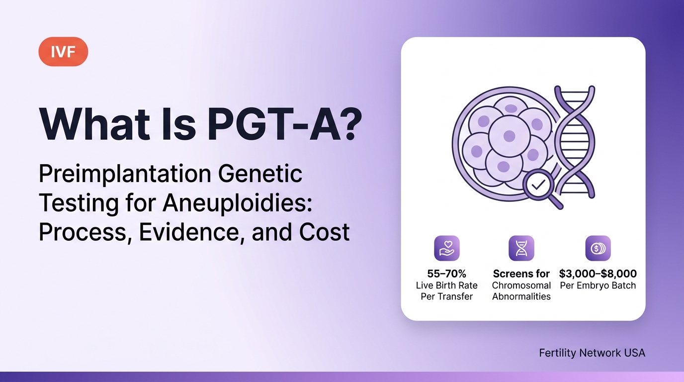
What is PGT-A?
PGT-A (preimplantation genetic testing for aneuploidies) is a chromosomal analysis performed on 5–10 cells biopsied from the trophectoderm (outer cell layer) of a day-5 or day-6 IVF blastocyst, using next-generation sequencing (NGS) to determine whether all 24 chromosome types are present in the correct copy number. The result classifies each embryo as euploid (chromosomally normal), aneuploid (chromosomally abnormal), or mosaic (mixed normal and abnormal cells).
PGT-A was formerly called PGS (preimplantation genetic screening). ASRM and PGDIS (Preimplantation Genetic Diagnosis International Society) adopted the standardized "PGT" terminology in 2017, replacing PGS with PGT-A, PGD with PGT-M, and chromosomal structural rearrangement testing with PGT-SR. The procedure is identical — only the name changed.
What is aneuploidy and why does it matter for IVF?
Aneuploidy is the presence of an abnormal chromosome number in an embryo — most commonly a missing chromosome (monosomy) or an extra chromosome (trisomy) — and is the leading cause of IVF implantation failure, early miscarriage, and chromosomal syndromes such as Down syndrome (trisomy 21). Aneuploid embryos either fail to implant, miscarry in the first trimester, or — in the case of viable aneuploidies — result in a live birth with a chromosomal syndrome.
Aneuploidy in IVF embryos is caused primarily by meiotic errors during egg production — specifically, errors in chromosome segregation during meiosis I and meiosis II as the egg matures. These errors increase with maternal age because the meiotic spindle becomes less accurate as eggs age. The rate of aneuploidy rises sharply after age 35 and becomes the dominant limiting factor in IVF success after age 38.
| Patient Age | Aneuploidy Rate of Blastocysts | Euploid Rate | Clinical Impact |
|---|---|---|---|
| Under 35 | 30 – 40% | 60 – 70% | Most embryos are euploid; PGT-A benefit is smaller |
| Age 35–37 | 45 – 55% | 45 – 55% | Roughly half of embryos aneuploid; PGT-A improves selection |
| Age 38–40 | 55 – 65% | 35 – 45% | Majority aneuploid; PGT-A significantly improves transfer efficiency |
| Age 41–42 | 70 – 75% | 25 – 30% | Most embryos aneuploid; PGT-A essential for identifying viable embryos |
| Over 42 | 75 – 85% | 15 – 25% | Very high aneuploidy; expect few euploid embryos per cycle |
How is PGT-A performed?
PGT-A is performed in 3 sequential steps: blastocyst trophectoderm biopsy by the embryologist, DNA amplification and next-generation sequencing (NGS) by the genetics laboratory, and results reporting and embryo selection before the frozen embryo transfer (FET) cycle. The entire process takes 1–2 weeks and means that PGT-A cycles always use frozen embryo transfer — never fresh transfer.
Blastocyst trophectoderm biopsy (Day 5 or Day 6)
On day 5 or 6 of embryo culture, the clinical embryologist performs a trophectoderm biopsy on each fully expanded or hatching blastocyst. The embryologist uses a laser to create a small opening in the zona pellucida, then removes 5–10 trophectoderm cells using a fine biopsy pipette and aspiration. The trophectoderm is the outer cell layer that develops into the placenta — biopsying it does not sample the inner cell mass (ICM), which becomes the fetus. Biopsied embryos are immediately vitrified. Cells are placed in a labeled tube and shipped overnight to the genetics laboratory.
Whole genome amplification + next-generation sequencing (NGS)
At the genetics laboratory, the DNA from the 5–10 biopsied cells undergoes whole genome amplification (WGA) — a PCR-based process that amplifies the tiny quantity of DNA from a few cells into sufficient material for sequencing. The amplified DNA is then sequenced using next-generation sequencing (NGS), which maps DNA reads across all 24 chromosome types to detect copy number variations. NGS has a resolution of approximately 10–20 Mb for segmental aneuploidies and detects all whole-chromosome gains and losses. Results are returned to the fertility clinic in 16–24 hours from the lab in most cases.
Results classification and embryo selection
Each embryo is classified into one of 3 categories: euploid (all 24 chromosome types in correct copy number — recommended for transfer), aneuploid (one or more chromosomes in abnormal copy number — not recommended for transfer), or mosaic (20–80% of cells aneuploid — lower priority for transfer; may be considered when no euploid embryos are available). The reproductive endocrinologist and patient review results and select the highest-priority euploid embryo for the FET cycle.
Who benefits most from PGT-A?
PGT-A provides the greatest clinical benefit for 4 patient groups: women over 37, patients with recurrent pregnancy loss (RPL), patients with recurrent implantation failure (RIF), and patients with a prior aneuploid pregnancy, according to ASRM Practice Committee guidelines (2023). For women under 35 with normal ovarian reserve, ASRM does not recommend routine PGT-A.
| Patient Profile | PGT-A Recommended? | Rationale |
|---|---|---|
| Women over 37 | Yes — strongly recommended | High embryo aneuploidy rate (55–75%); PGT-A identifies viable embryos efficiently |
| Recurrent pregnancy loss (RPL) (2+ miscarriages) | Yes — recommended | 50–60% of recurrent miscarriages are caused by embryo aneuploidy; PGT-A reduces recurrence |
| Recurrent implantation failure (RIF) (3+ failed transfers of morphologically normal embryos) | Yes — recommended | Identifies aneuploid embryos that appeared morphologically normal; improves transfer efficiency |
| Prior aneuploid pregnancy (trisomy 21, 18, 13, or other) | Yes — recommended | Prior aneuploidy increases recurrence risk; PGT-A screens future embryos before transfer |
| Women 35–37 with good ovarian reserve | Yes — consider | Aneuploidy rate 45–55%; benefit is significant especially if only a few blastocysts |
| Women under 35 with normal reserve, no prior failure | No — ASRM does not recommend routine | Aneuploidy rate 30–40%; most embryos euploid; risk of discarding viable mosaic embryos outweighs benefit |
| Single embryo transfer strategy (any age) | Yes — improves SET confidence | Confirming euploidy before single embryo transfer maximizes per-transfer success rate |
What live birth rates does PGT-A achieve per euploid embryo transfer?
Transferring a PGT-A-confirmed euploid blastocyst achieves a 55–70% live birth rate per transfer regardless of the patient's age, because euploid embryos fail primarily due to uterine and implantation factors rather than chromosomal abnormality. This is the key clinical argument for PGT-A in older patients — a 41-year-old's euploid embryo achieves a similar per-transfer rate as a 32-year-old's euploid embryo.
| Patient Age | Live Birth Rate / Transfer Without PGT-A | Live Birth Rate / Transfer With PGT-A (Euploid Only) | Per-Transfer Improvement |
|---|---|---|---|
| Under 35 | 51.2% | 60 – 70% | +9 – 19 percentage points |
| Age 35–37 | 38.6% | 58 – 68% | +20 – 30 percentage points |
| Age 38–40 | 27.8% | 55 – 65% | +27 – 37 percentage points |
| Age 41–42 | 16.3% | 50 – 60% | +34 – 44 percentage points |
| Over 42 | 8.1% | 45 – 55% | +37 – 47 percentage points |
What is a mosaic embryo in PGT-A?
A mosaic embryo contains a mixture of euploid and aneuploid cells — detected by NGS when 20–80% of the trophectoderm biopsy cells show chromosomal abnormalities. Mosaic embryos occupy a middle category between fully euploid (recommended for transfer) and fully aneuploid (not recommended). The proportion of abnormal cells and which chromosomes are affected determine the clinical priority of a mosaic embryo.
Mosaic embryos can produce healthy live births: published data shows a 35–50% live birth rate per mosaic embryo transfer— lower than euploid (55–70%) but significantly higher than untested embryos in older patients (8–27%). The PGDIS (Preimplantation Genetic Diagnosis International Society) 2023 guidelines recommend considering mosaic embryo transfer when:
| Mosaic Category | Definition | Transfer Priority | Live Birth Rate / Transfer |
|---|---|---|---|
| Euploid | 0–20% abnormal cells | Highest — transfer first | 55 – 70% |
| Low-level mosaic | 20–40% abnormal cells | Second — transfer if no euploid available | 45 – 55% |
| High-level mosaic | 40–80% abnormal cells | Third — transfer with counseling | 35 – 45% |
| Aneuploid | 80–100% abnormal cells | Not recommended for transfer | Less than 5% |
Mosaic embryo transfer requires additional genetic counseling and informed consent. Prenatal testing — either NIPT (non-invasive prenatal testing) or amniocentesis — is recommended in all pregnancies resulting from mosaic embryo transfer to confirm fetal chromosomal status. Outcomes reported from mosaic embryo transfers show that the large majority of live births from low-level mosaic embryos are chromosomally normal — the mosaicism appears to self-correct during development, with euploid cells selectively populating the fetal lineage.
What is the difference between PGT-A and PGT-M?
PGT-A screens embryos for chromosomal copy number errors (aneuploidies); PGT-M screens embryos for a specific single-gene (monogenic) disease mutation that one or both parents are known to carry. PGT-A and PGT-M are distinct tests, require different laboratory methods, and serve different clinical indications. They can be performed simultaneously on the same embryo biopsy sample.
| Factor | PGT-A | PGT-M |
|---|---|---|
| What it detects | Chromosomal copy number errors (monosomy, trisomy) | Specific single-gene mutations (BRCA1, CFTR, HTT, SMN1, etc.) |
| Who it is for | All IVF patients meeting age or clinical criteria | Known carriers of a single-gene disease (autosomal dominant, recessive, or X-linked) |
| Laboratory method | Next-generation sequencing (NGS) copy number analysis | Custom probe design + NGS or PCR; requires 6–8 weeks of probe development before cycle |
| Setup time required? | No — off-the-shelf test; results in 16–24 hours | Yes — 6–8 weeks for custom probe development before first cycle |
| Cost | $3,000 – $8,000 per batch | $6,000 – $12,000 per batch (higher due to custom probe development) |
| Common indications | Age over 37, RPL, RIF, prior aneuploid pregnancy | BRCA1/2 carriers, cystic fibrosis, Huntington's disease, spinal muscular atrophy |
A third test category — PGT-SR (structural rearrangements) — screens embryos from parents who carry balanced chromosomal translocations or inversions. PGT-SR detects unbalanced chromosomal configurations that would cause recurrent miscarriage even with morphologically normal embryos. PGT-SR is indicated for patients with confirmed reciprocal or Robertsonian translocations.
How accurate is PGT-A?
PGT-A using next-generation sequencing (NGS) achieves a diagnostic accuracy of 97–99% for whole-chromosome aneuploidies and 95–98% for segmental aneuploidies larger than 10 Mb, based on validation studies comparing NGS results against full karyotype of the same embryos. The remaining 1–3% error rate represents both false positives (euploid embryos classified as aneuploid) and false negatives (aneuploid embryos classified as euploid).
| Error Type | Rate | Clinical Consequence |
|---|---|---|
| False positive (euploid embryo classified as aneuploid) | 1 – 3% | Viable embryo not transferred; missed opportunity |
| False negative (aneuploid embryo classified as euploid) | Less than 1% | Aneuploid embryo transferred; higher miscarriage risk or affected pregnancy |
| Inconclusive result (amplification failure; insufficient DNA) | 2 – 5% | No result for that embryo; embryo remains in storage pending re-biopsy decision |
| Mosaic misclassification (mosaic classified as euploid or aneuploid) | Variable | Depends on direction of misclassification; prenatal testing post-transfer recommended |
The false positive rate is the most clinically significant limitation of PGT-A for younger patients. A 1–3% false positive rate means that 1–3 embryos per 100 analyzed are discarded despite being chromosomally normal. For a 32-year-old patient with 8 euploid embryos out of 10 blastocysts, this risk is small in absolute terms. For a 40-year-old with only 2 blastocysts — both potentially mosaic or borderline — a false positive result could mean no embryos available for transfer in that cycle.
Does PGT-A improve IVF outcomes for all patients?
PGT-A improves per-transfer live birth rates for all age groups, but does not improve cumulative live birth rates per initiated cycle for women under 35 with normal ovarian reserve — and ASRM explicitly does not recommend routine PGT-A for this population. This is the central clinical controversy around PGT-A.
The evidence comes from 2 landmark randomized controlled trials: the STAR trial (Scott et al., 2013) showed PGT-A significantly improved outcomes for women aged 35–42, while the RCT by Munné et al. (2019) showed improved per-transfer rates across all ages but no improvement in cumulative live birth rates per cycle for women under 35. A 2022 Cochrane review concluded that for women under 35 with good prognosis, PGT-A may increase per-transfer success but does not improve overall live birth rates because it reduces the number of embryos available for transfer by eliminating aneuploid and mosaic embryos.
How much does PGT-A cost?
PGT-A costs $3,000 to $8,000 per embryo batch tested, including the trophectoderm biopsy performed by the embryologist and the NGS analysis performed by the genetics laboratory. The total cost depends on the number of embryos biopsied — most labs charge a per-embryo fee for biopsy and a per-batch fee for sequencing.
| PGT-A Cost Component | Average Cost | Notes |
|---|---|---|
| Trophectoderm biopsy (per embryo) | $200 – $500 per embryo | Performed by clinical embryologist; billed per embryo biopsied |
| NGS laboratory analysis (per batch) | $1,800 – $3,500 per batch | Fixed batch fee for 1–8 embryos; charged per batch sent to lab |
| Genetic counseling session | $200 – $500 | Recommended before PGT-A; sometimes included in clinic fee |
| Total PGT-A (2 embryos biopsied) | $2,200 – $4,500 | 2 biopsy fees + 1 batch sequencing fee |
| Total PGT-A (4 embryos biopsied) | $2,600 – $5,500 | 4 biopsy fees + 1 batch sequencing fee |
| Total PGT-A (6–8 embryos biopsied) | $3,000 – $7,500 | May span 2 batch fees depending on timing |
PGT-A is covered by fertility insurance mandates in several states when medically indicated — including for recurrent pregnancy loss and prior aneuploid pregnancy — but coverage for routine PGT-A based on age alone is inconsistent. FSA and HSA funds can be used for PGT-A costs. For the full IVF cost breakdown including PGT-A, see IVF Cost in the USA: Average Price Per Cycle, by State, and Hidden Fees.
Frequently Asked Questions
Can PGT-A detect Down syndrome?+
Yes — PGT-A detects trisomy 21 (Down syndrome) with 97–99% accuracy, along with all other whole-chromosome aneuploidies including trisomy 18 (Edwards syndrome), trisomy 13 (Patau syndrome), monosomy X (Turner syndrome), and sex chromosome aneuploidies (XXY, XYY, XXX). Embryos with these chromosomal abnormalities are classified as aneuploid and are not recommended for transfer. Prenatal testing (NIPT or amniocentesis) remains recommended after PGT-A even in confirmed euploid pregnancies, given the 1–3% false negative rate and the fact that PGT-A does not screen for single-gene disorders.
Does PGT-A reduce miscarriage risk?+
PGT-A reduces miscarriage rate per embryo transfer from 15–40% (untested embryos, age-dependent) to 5–10% (euploid embryos) by eliminating chromosomally abnormal embryos before transfer, according to multiple large prospective cohort studies. The miscarriage reduction is most significant for women over 38, where 50–70% of miscarriages in untested IVF cycles are caused by embryo aneuploidy. For women under 35, the absolute miscarriage reduction is smaller — approximately 5–8 percentage points — because baseline aneuploidy rates are lower and other miscarriage causes (uterine, immunological, thrombophilic) represent a larger proportion of total losses.
Does PGT-A damage the embryo?+
Trophectoderm biopsy for PGT-A does not damage the embryo's developmental potential when performed on fully expanded day-5 or day-6 blastocysts by an experienced embryologist. The biopsied cells are trophectoderm — future placenta — not inner cell mass (future fetus). Multiple large-scale outcome studies (SART data analysis; Belgian VUB 10-year follow-up) show no difference in obstetric outcomes, birth weight, or child development between children born from biopsied and non-biopsied embryos. The 1–5% embryo loss rate attributable to biopsy (vitrification failure after biopsy, or embryo arrest during biopsy) is the primary procedural risk, not fetal developmental harm.
How is PGT-A different from prenatal testing (NIPT or amniocentesis)?+
PGT-A tests the embryo before uterine transfer; NIPT and amniocentesis test a developing pregnancy after implantation. PGT-A allows chromosomally abnormal embryos to be excluded before any pregnancy is established, avoiding the need for pregnancy termination. NIPT tests cell-free fetal DNA from maternal blood at 10+ weeks of pregnancy and detects the most common aneuploidies (trisomy 21, 18, 13) with 97–99% sensitivity but cannot test all 24 chromosomes with equal accuracy. Amniocentesis tests amniotic fluid cells at 15–20 weeks and provides a full fetal karyotype but carries a 0.1–0.3% procedural miscarriage risk. PGT-A and prenatal testing serve complementary roles — ASRM recommends prenatal testing even after PGT-A, given the 1–3% PGT-A false negative rate.
Can aneuploid embryos be re-tested if the result seems wrong?+
Aneuploid embryos can be re-biopsied and re-tested at some specialized centers, but re-testing is not routinely recommended by ASRM or PGDIS because repeated biopsy further reduces the cell number available in the inner cell mass and trophectoderm, increasing the risk of embryo damage. A small number of centers offer "rescue biopsy" programs for patients with no euploid embryos — re-testing mosaic or borderline aneuploid embryos to confirm the result before discarding them. Re-test concordance rates for whole-chromosome aneuploidies are approximately 92–95%, meaning 5–8% of confirmed aneuploid results reclassify on re-test. For patients with no euploid embryos and at least one mosaic embryo, transferring the best-quality mosaic embryo (after genetic counseling) is generally preferred over embryo re-biopsy.
Fertility Network USA is an independent information directory. All PGT-A clinical guidance, aneuploidy rates, live birth rates, and accuracy data in this article are sourced from ASRM Practice Committee guidelines (2023), PGDIS position statements (2023), and peer-reviewed reproductive genetics literature including Cochrane systematic reviews. PGT-A accuracy figures represent published NGS validation study ranges. Individual outcomes vary significantly by genetics laboratory, embryo quality, patient age, and clinical indication. PGT-A results do not guarantee a healthy pregnancy — prenatal testing remains recommended after any PGT-A cycle. This article does not constitute medical advice. Always consult a board-certified reproductive endocrinologist and genetic counselor before pursuing PGT-A.